
Super User
Equine Deworming Information
AAEP’s Parasite Control Guidelines
AAEP’s Parasite Control Guidelines
Commonly used strategies for parasite control in adult horses are based largely on knowledge and concepts that are more than 50 years old. However, much has changed in this time, necessitating a re-examination of recommendations for parasite control.
In response to this need, the AAEP’s Parasite Control Subcommittee of the Infectious Disease Committee in 2013 produced a comprehensive set of recommendations for helping veterinarians develop improved strategies and programs for parasite control in horses of all ages. In 2019, these guidelines went through a rigorous review with the committee and former subcommittee and were updated.
It is important to keep in mind that the information contained within these guidelines are suggestions; there are many variations of these suggested programs that will still meet the same goals and follow the same principles. Ultimately, each farm (with veterinary guidance) should develop its own program tailored to the specific needs of the farm and each animal. There is no such thing as a “one size fits all” program.
Guidelines are specified separately for adult and young horses (less than 3 years). All treatment and non-treatment recommendations are made within the context of a preventive program where fecal egg count (FEC) surveillance is being performed.
Read Parasite Control Guidelines.
Visit AAEP's Parasite Control Guidlines webpage
Equine Deworming Plan
Equine Deworming Plan
The recent increase in parasite resistance to commonly used deworming products has left the equine community concerned and in search of ways to slow down, if not reverse, the alarming trend. As parasites can pose a very serious threat to a horse’s health, including fatal situations, it is apparent that the commonly accepted approach to parasite control should be reconsidered. For example, the so called dewormer “rotation” is no longer an acceptable practice.
Strongyles, roundworms, bots, pinworms, and tapeworms are some of the parasites that veterinarians and horse owners deal with. Some of these can be identified easily, others not so much. That is why every treatment should start with analyzing fecal samples. The test is relatively inexpensive, and can provide a solid basis for choosing the right product that will ensure an effective deworming treatment. Every horse from the herd should be tested, but if the group is very large, then a proportionate test group may be used to gather samples from. The fecal test should be done once a year or at least every other year.
Statistically, horses can be categorized as follows:
| Number of eggs in a gram of fecal matter (EPG) | Category | Proportion relative to the herd |
|---|---|---|
| More than 600 (up to 3,000) | Heavy shedder | 20% |
| 200 - 500 | Moderate shedder | 30-40 % |
| 0 - 100 | Light shedder | 40-50 % |
If you test the whole herd individually you may find that not all horses carry the same amount and/or type of parasites. Some horses will carry a lot of worms and others much less. This is due to individual immunity, genetics, exposure level, and a variety of other factors. It may be a good idea to group horses based on the parasite type of infestation they have. This may help prevent further transfer of eggs and larvae between individuals.
There is no longer any fixed time chart or an ideal schedule for equine deworming. Instead, a proper deworming program will be based on the level of infestation and on the climate in your area. It is strongly recommended to perform a treatment after the first killing frost in the late fall to control bots and another round in the spring with a praziquantel- containing dewormer, which will provide a solid tapeworm control program. Note that this is the minimal treatment for the so-called light shedders.
| Category | Recommended frequency of deworming per year |
|---|---|
| Light shedder | 2 times per year (or no treatment at all) (late fall after the first killing frost and spring) |
| Moderate shedder | 3 – 5 times per year (late fall after the first killing frost and spring , plus when needed, usually in the summer season in the warm environments) Will benefit from one additional treatment during the main season of pasture transmission –spring through autumn in the North; autumn through spring in the South* |
| Heavy shedder | 5-6 times per year (approximately every other month) Intensive treatment required all through the main seasons of transmission* |
Always consult your veterinarian before treatment. Fecal testing can help determine the right product and the most effective way to treat your horses. The currently preferred drugs are pyrantel (not effective for tapeworm or bot treatment), ivermectin, moxidectin, and praziquantel (effective against tapeworm).
*Craig R. Reinemeyer, DVM, PhD, The Horse.com, “What is your Horse’s Fecal Egg Count Telling You?”, April 17, 2017
Resistance
Resistance
Resistance is the ability of worms in a population to survive treatment by a particular deworming chemical. Resistance grows when parasites survive treatment, and then pass the ability to survive on to their offspring.
Factors that influence resistance growth1:
- Under dosing.
- Deworming frequency.
- Using dewormers that do not kill resistant parasites.
Resistance can be reduced by:
- Use the proper dose and ensure the horse ingests the full dose.
- Deworm only when needed.
- Use dewormers that are effective. Studies have shown certain deworming classes, e.g. benzimidazoles have a high rate of resistance2.
80% of horses in a recent study3 whose weights were set visually were underestimated. Optimally, weigh your horse. This can be done when trailering, weigh your vehicle with your horse and without (scales can be found at feed mills, truck stops etc). If a scale is unavailable, weight tapes use a combination of a horse’s girth and height to estimate weight. Use care, weight tapes can be inaccurate for foals, miniatures and heavily muscled and high withered horses.
Know how many pounds the dewormer treats. This varies by brand, by as much as 20%. Round up to the nearest measurement and securely lock the syringe mechanism in place for that dosage.
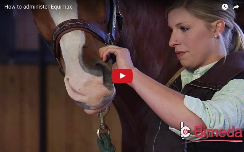
Be sure once administered the horse ingests the full dose. This can be aided by raising the horses head and stroking under their jaw. Apple flavored dewormers can increase the ease of dosing, this can help reduce spit outs and other activities that prevent a full dose from being ingested.
- Sangster NC. Pharmacology of anthelmintics resistance in cyathostomes: will it occur with the avermectin/milbemycins? Vet. Parasitol. 1999; 85: 189-204.
- Kaplan RM. Anthelmintic resistance in nematodes of horses. Vet. Res. 2002; 33: 491-507.
- Asquith, R. Johnson, E. Kivipelto, J. and Depew, C. (1990). Erroneous weight estimation of horses. Proceedings of the annual convention of the American Association of equine practitioners. 599-607.
Guarantee
We stand behind our products, with the
Bimeda 100% Dewormer Satisfaction Guarantee
No-hassle satisfaction guarantee when you treat your horse according to label directions with any of Bimeda’s equine dewormers.
Return Instructions
If you are not satisfied with the level of parasite control that you received:
- Call Bimeda customer service at 888-524-6332 to obtain a guarantee claim number.
Satisfaction Guarantee claims must be made within one (1) month of administration and six (6) months of purchase. - Submit your itemized printed receipt for the purchase of Equimax®, Bimectin® or Exodus® Branded Products, date of product administration, product package and your Satisfaction Guarantee claim number.
- Your request will be reviewed and if all requirements are met, you will receive a product replacement or refund.
NOTE: Bimeda reserves the right to cancel or modify this guarantee at any time. Void where prohibited by law or regulation. All federal, state and local laws and regulations apply. Guarantee valid for horse owners only. Bimeda customers, Veterinarians, retailers and distributors do not qualify for this guarantee.
Bimeda's Line of Dewormers


Tapeworms - A serious threat to horses in the U.S.

Recent research revealed that more than 50% of horses tested in the United States have been infected with tapeworms.
Thanks to increased awareness and the availability of EQUIMAX® to treat tapeworms, you can take steps to reduce your horse´s risk of illness associated with this prevalent parasite.
Ileal Impaction Colic
- Occurs when an obstruction prevents the passing of digested material.
- Research indicates that more than 80% of these colics are associated with tapeworms.2
EQUIMAX® controls tapeworms (A. perfoliata) that may cause certain types of colic.
Inside the Tapeworm
Tapeworm Species
- Anoplocephala perfoliata
- Anoplocephala magna
- Paranoplocephala mamillana
Three species of equine tapeworm affect the horse. Anoplocephala perfoliata is by far the most prevalent and is of the most concern to horse owners. Anoplocephala magna and Paranoplocephala mamillana, however, do occur elsewhere in the world but are rare in the United States.
Tapeworm Characteristics
The tapeworm belongs to a class of parasites known as cestodes - unlike roundworms and strongyles, which are nematodes. Tapeworms have a simple body structure, composed of a scolex, which attaches to the intestinal wall of the horse, and many proglottids, or body segments, each of which contains reproductive organs and eggs. The tapeworm receives nutrients through its tegument - its absorptive outer layer.
Anoplocephala perfoliata

Anoplocephala perfoliata is the most abundant and infective of the equine tapeworms. It usually measures about 1 to 3 inches in length and has a rounded scolex with four hooks that allow it to attach to the horse’s intestin.
Tapeworm Eggs
The very nature of tapeworm eggs makes diagnosis of their presence extremely difficult. Tapeworm eggs occur in very small numbers, and many times they exist in packets rather than as individual eggs. The excreted tapeworm eggs don't float well in traditional fecal floatation tests, which means they evade veterinary detection quite easily.
The Ileocecal Junction
Anoplocephala perfoliata tend to congregate on the surface of a unique area in the horse - the ileocecal junction. This spot is the common opening between the ileum, or small intestine, the colon and the cecum, the pouch that is the beginning of the large intestine and serves as a kind of fermentation vat.
To get a better understanding of the tapeworm's life cycle and how it affects the horse, let's take a look at the anatomy of a normal horse Anatomy of a normal horse.
An Indirect Life Cycle
All tapeworms have an indirect life cycle; they need an intermediate host in order to complete their life cycle. Equine tapeworms use an extremely common, tiny, pasture-dwelling insect for this purpose - the oribatid mite. These mites are very small, and they're found in virtually all soils all over the world.
Lifecycle of the Tapeworm
- Oribatid mites are invisible to the eye and can be found on many sources such as hay, feedstuffs, straw and pastures.
- Tapeworm-infected horses may appear in good health and do not exhibit visible signs of damage, such as dull hair coat and diarrhea.
- Because tapeworm eggs are contained in packets and do not float well, fecal egg tests are only 3.1% accurate, making it extremely difficult to diagnose tapeworms in affected horses.3
- Left untreated, tapeworms can cause serious health problems and result in death.
Tapeworm Damage
Tapeworm Colonization
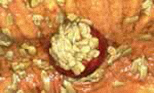
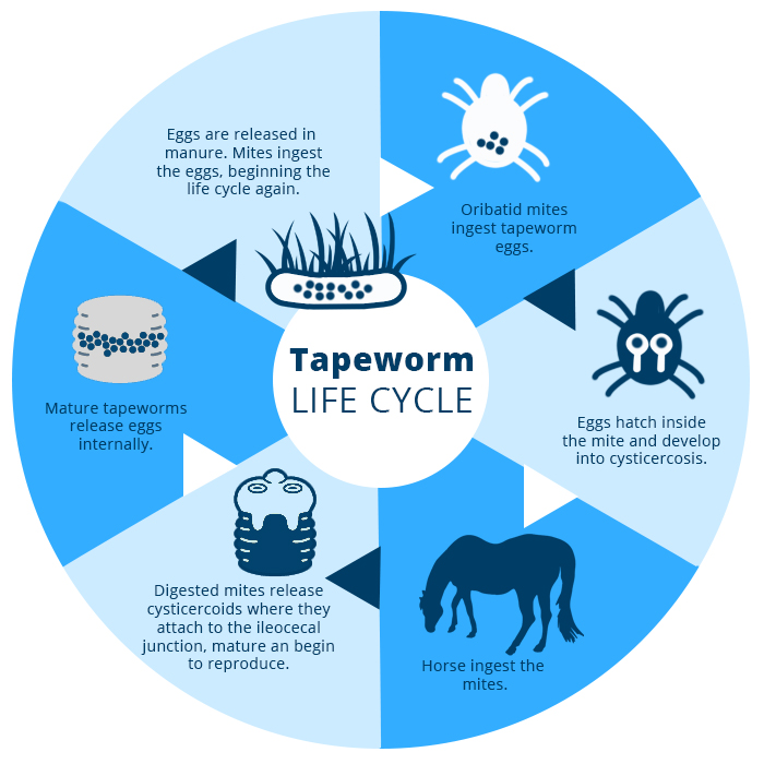
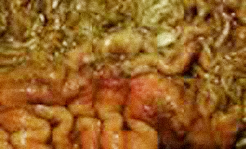
So why is it important that so many horses are exposed to tapeworms? What kind of damage do tapeworms cause that makes them such a threat? The damage begins because of the way that Anoplocephala perfoliata colonize around the ileocecal junction. As this image shows, the large numbers of parasites concentrated in a single area, and the fact that they attach to the mucosa with strong hooks, create a tremendous amount of irritation and inflammation at this important junction of the small intestine, cecum and colon. Let's watch as this occurs Tapeworm Inflammation.
Tapeworm Attachment
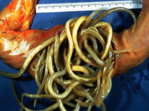
Tapeworms attach tightly to the gut wall. This picture illustrates that attachment and shows the ulcerations it can leave behind.
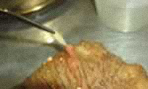
Damage to the Gut Wall
Tapeworms can spread out over a fairly large area around the ileocecal junction if the infection is severe enough. Their presence causes irritation wherever they go. This section of gut wall has had a number of tapeworms removed from it, revealing the damage their presence creates.
Ileocecal Intussusception
The tapeworm has been indicated in three specific types of colic in horses. The first is intussusception. Remember that ingested material is moved through the digestive tract with the help of waves or peristalsis. When tapeworms are present, the inflammation they cause can exaggerate these waves at the ileocecal junction, and the small intestine may actually push its way into the cecum as a result. This type of colic is very serious, and virtually all intussusception colics are caused by tapeworm infections. Let's take a closer look Ileocecal Intussusception.
Impaction Colic
The disruption of normal peristalsis through the ileocecal junction can cause impaction colic. In ileocecal impaction colic, fecal matter in the cecum isn't expelled and backs up into the small intestine. This is a long-term, slow colic, as additional digested materials continue to pile up. Here's how it works Impaction Colic.
Spasmodic Colic
Spasmodic or gas colic is extremely common in horses and often associated with tapeworms. It is caused by gas trapped as a result of abnormal peristalsis. The peristaltic waves become erratic, and the intestine seems almost to spasm. This is what it looks like Spasmodic Colic.
Tapeworm Damage Summary
Tapeworms are a threat to the health of horses in the United States and around the world. This pest causes severe inflammation at the ileocecal junction and affects normal peristalsis of the gut, impairing a horse's condition and performance, while causing multiple types of potentially life-threatening colics.
The correlation between tapeworm presence and the occurrence of colic is strong. Tapeworms are the primary cause of all ileoccecal intussusceptions. Tapeworms cause approximately 81 percent of ileocecal impaction colics and 22 percent of spasmodic colics.
Now that we know the incidence of tapeworm exposure and the significant damage these parasites cause, what can owners do to protect their horses from this threat?
1. C.R. Reinemeyer, A.W. Farley, S.A. Kania, B.W. Rohrbach and R.H. Dressler, 48th Annual Meeting of the American Association of Veterinary Parasitologists, Denver, CO, July 2003.
2. C.J. Proudman, N.P. French and A.J. Trees, Equine Veterinary Journal 1998; 30 (3): 194–199.
3. E.T. Lyons, S.C. Tolliver, J.H. Drudge et al. Parasites in Kentucky Thoroughbreds at necropsy: Emphasis on stomach worms and tapeworms. American Journal of Veterinary Research 1993; 44: 839–844.
4. Mercier P., Bousquet E., Sanquer A, et al. Time of re–appearance of fecal tapeworm eggs in horses treated with an ivermectin–praziquantel combination and left in their contaminated environment. World Association Adv. Vet Parasitol 21st International Conference. 19–23 August, 2007, Ghent, Belgium.
Tapeworms

Much uncertainty surrounds the significance of tapeworm infections in horses. The natural infection is hard to diagnose in the living animal, and no successful experimental infections have been reported.
The equine tapeworm was considered for many years to be an incidental finding in the intestinal tracts of horses at postmortem examination and rarely associated with clinical disease. Infections usually went undetected because of the difficulties of fecal egg examinations and because no clearly defined clinical symptoms were associated with tapeworms.
Recent studies, however, have defined this parasite as a significant risk factor for spasmodic colic and impactions at the terminal end of the small intestine. Add to this several studies showing tapeworm infection rates as high as 81.5 percent, and an interesting story emerges about a parasite that was previously considered harmless.
Tapeworm Species
Three species of tapeworms are known to infect the horse. Anoplocephala perfoliata, Anoplocephala magna and Paranoplocephala mamillana. Studies have demonstrated A. perfoliata to be the most common tapeworm in the horse. A. magna is the next most common, and P. mamillana is quite rare.
A. perfoliata is a short, yellow/green tapeworm with a triangular body. The size of mature worms varies from three to eight centimeters – tapeworms smaller than this are either immature worms or P. mamillana. The head of A. perfoliata is equipped with four suckers with which the parasite can secure itself to the mucosa of its host. Nutrients are absorbed through the parasite’s cuticle.
Tapeworm Diagnosis
Diagnosing tapeworm infection is difficult. Most infections have no symptoms, but cause subclinical damage that may be present for some time before causing visible disease. One textbook describes unthriftiness and anemia as consequences of heavy infestation, but moderate infections may go unnoticed.
Another factor making diagnosis difficult is the nature of tapeworm eggs. They occur in very small numbers and often in packets rather than individual eggs. They don’t float well in the testing medium, so they are often missed in routine fecal egg-count analysis. More sophisticated tests are needed to visualize tapeworm eggs. Most veterinary practices and labs aren’t equipped to do these tests, so, in many cases, the diagnosis is missed. Sometimes, however, tapeworm specimens can be found in the feces of infected horses after they have been treated with drugs active against these parasites.
The Tapeworm Life Cycle
The tapeworm is different from many other parasites because it has an indirect life cycle. This means it requires an intermediate host in order to develop. The intermediate host of A. perfoliata is the free-living oribatid mite. This mite is found in very large numbers on pastures and often even in hay and straw.
Inside the mite, the tapeworm egg undergoes cellular division and development to become a larva. This process takes 12-15 weeks. Inadvertent ingestion of mites containing infective larvae occurs as horses graze, and it can result in tapeworm infection. Larvae then develop within the primary host – the horse – to mature tapeworms. They can begin shedding segments full of eggs in 6-10 weeks. There appears to be little seasonal variation in the number of horses infected with tapeworms or in the magnitude of infection.
The equine tapeworm usually congregates around the ileocaecal valve in the intestinal tract, at the junction between the large and small intestines. It is in this small area of the horse’s intestinal tract that changes attributable to tapeworms are found. Until recently, few studies had been done of the damage caused by tapeworms.
Tapeworm Damage
The tapeworm can cause severe damage inside the horse’s intestinal tract. Its presence can lead to decreased intestinal motility and colic.
Until the life cycle of A. perfoliata – the common tapeworm – is better understood, it is difficult to decide when to provide horses with appropriate anti-tapeworm drugs. Generally, treatments directed against tapeworms should be given every six months, with treatment in the fall and again in late spring. The role of tapeworms in equine colic should be kept in perspective. They represent a small, but avoidable, risk in certain types of colic.
Roundworms (Parascaris equorum)
The roundworm, or ascarid, is a prolific egg layer. Each female can lay from 100,000 to 200,000 eggs each day. The eggs pass out of the horse with the feces. Infective larvae develop within the eggs, which are triple-coated and are not affected by adverse weather conditions. Therefore, they remain viable for up to 10 years. It is important to remember that fecal tests do not detect migration of parasite larvae within the horse.
Ascarid Eggs
This is an ascarid egg, which may have been lying on the ground for 10 years. When it is ingested, it begins an amazing journey. The egg’s coating is digested in the stomach. As the eggs reach the small intestine, they hatch, and the larvae immediately penetrate the lining of the intestinal tract, beginning a 30-day migration.
Liver With Ascarid Larvae Damage
The eggs travel via the hepatic vein to the liver, where they eat their way around the liver for seven to 10 days. The real damage takes place here and in the lungs. Fortunately, the liver is a very resilient organ and can regenerate itself. We seldom see any permanent damage to the liver from ascarid larval migration.
Small Hemorrhages From Ascarid Larvae Migration
The larvae then go to the lungs and continue their migration for 14-21 days, again eating their way around lung tissue. Damage done to the lungs is a different story than that of the liver, because the lungs, which heal by scarring, do not regenerate. This damage is permanent. After the ascarids mature and are ready to complete migration, they burrow from the blood side of the lung into the air side.
When migrating ascarid larvae are present, the immune system reacts violently to the foreign protein and destroys the alveoli. Such damage predisposes foals to pneumonia and may result in pulmonary hemorrhaging in a horse that becomes an athlete. When a horse is just a few months old, it has all the lung tissue it is ever going to have. Because lung tissue heals by scarring, damage to these sensitive structures is permanent. There will be less functional lung for the horse to use.
Horses whose lungs have been damaged by ascarid larval migration may have to breathe harder and faster to meet their oxygen demands as they develop and are asked to perform.
The worms then crawl from the alveoli into the bronchioles, to the bronchi, and into the trachea. They cause enough irritation to elicit a cough, so they are coughed to the back of the throat and re-swallowed as mature larvae. As adults, they swim upstream in the small intestine, robbing the horse of nutrition. These parasites have a very efficient migration. When the larvae reach the small intestine for the second time, their presence causes little consequence to the horse.
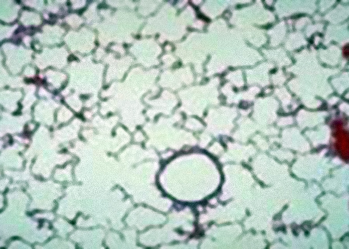 Normal Lung Tissue, This microscopic look at normal lung tissue shows empty air spaces – the alveoli – where oxygen is exchanged for CO2.
 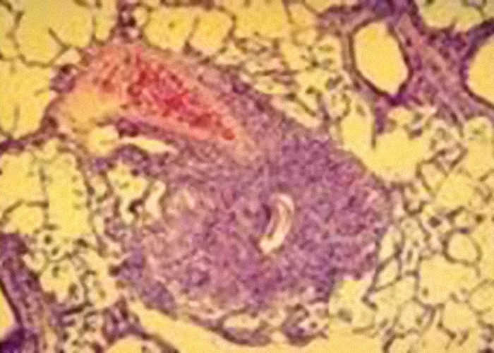 Damaged Lung Tissue, This image shows lung tissue damaged by ascarid larval migration.
 Ascarid Larval Migration
Although horses may develop an immunity to ascarids, that immunity does not prevent the migration and damage these parasites may cause.
Ascarid larval migration can lead to other diseases. It reduces overall thriftiness in foals and can be related to foalhood pneumonia. Ascarid larvae may have an immunosuppressive effect in the lungs, which means they can reduce the immune system’s ability to respond to foreign invaders like bacteria and viruses. Remember, fecal tests can’t detect these migrating larvae.
Horses Live in Contaminated Areas
Think for a moment about how these roundworm survival attributes could affect your horses. The ascarids in your horses are still producing hundreds of millions of eggs. The eggs are not affected by climatic conditions, and they survive for up to 10 years. The only difference between your horses and horses in a natural setting is that your horses don’t migrate 25 miles a day away from these eggs. Instead, they are contained in small grazing areas, which ensures that they ingest parasites. Parasitism has become a chronic disease that horses are faced with daily.
Pinworms (Oxyuris equi)
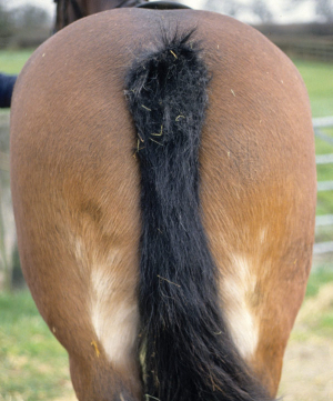
Pinworms have the most efficient life cycle of all the parasites that infect the horse. They don't migrate through any organ tissue, and they have developed a means of reproduction by which the eggs don't leave the herd of horses.
While the horse is relaxed or sleeping, female pinworms crawl out of the horse's rectum, deposit eggs and a sticky substance on the perianal region of the horse, and crawl back into the rectum. Infective pinworm eggs are ingested orally and, once in the colon, the larvae develop through various stages before becoming sexually mature in about five months. As horses migrate, they take the eggs and adults with them.
Anal Irritation From Pinworms
Fortunately, about the only damage that pinworms cause is itching of the tail head. This annoys the horse but doesn't threaten its life. Because pinworms spend their entire lives in the lumen of the intestine and don't migrate, they cause very little physical damage to the horse. Horses can have massive pinworm infections without exhibiting significant health problems.
What To Look For
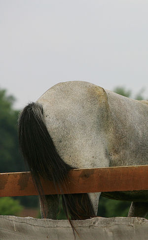 Keep an eye out for skin irritation around the anus area, rubbing of the tail, as well as biting and licking of hindquarters. In some situations, changes in behavior such as nervousness and loss of appetite may occur. Due to their life cycle, fecal tests are not a reliable method for detecting pinworm infestation. Veterinarians sometimes use the “scotch tape test”. This method uses tape that is pressed on the anal area and is then examined under a microscope.
The typical infected horse will go in search of places to rub their bottom in an effort to relieve the itching. When a horse scratches itself, residue from this sticky sub- stance can be left on surfaces. The larvae can also drop into the paddock or stable floor. Because of these circum- stances, horses are vulnerable to becoming infected and/or re-infected for up to four (4) months.
What to do?
Pinworm infestations are not easy to get rid of. Good stable management practice is needed, in addition to the proper dewormer.
For your horse - thoroughly cleanse the anus/tail and other affected areas before ad- ministering an appropriate dewormer.
For the stable - disinfect grooming kits, water and feed buckets, as well as other items the horse has rubbed on or come into contact with.
Strongyles
While pinworms and roundworms have fairly efficient life cycles, strongyles have a very unusual method of reproduction, especially considering that they developed in a desert environment. Large and small strongyles share identical life cycles outside of the horse but behave very differently once they're ingested.
Eggs are passed in the fecal material, just like ascarid eggs. Unlike ascarid eggs, which remain in egg form until they are ingested by the horse, strongyle eggs hatch into larvae in the manure paddy when weather conditions are favorable. From here, they go through a series of three molts. It's the third stage that can infect the horse. Under optimal conditions, third-stage larvae can live for up to three months. For their species to survive, strongyles must be ingested shortly after molting to this stage.
Strongyle Larvae in Dew Drop
To increase its chances for survival, the larva climbs up on blades of grass where a horse can ingest it while grazing. This is less likely under natural conditions, because the horse herd that deposited this larvae-ridden manure may have migrated many miles.
Strongyles are prolific egg layers. A single horse can pass 75-100 million eggs daily. The eggs can crawl up and down a blade of grass many times or bury themselves in the soil to protect against adverse weather.
Small Strongyles
Small strongyles also developed another unique trait - the ability to encyst themselves in the horse's tissues and remain in an encysted state for a prolonged time. Unfortunately, it causes real problems for the horse.
This encysted state helps the small strongyle: When larvae are ingested, they immediately penetrate the bowel lining and begin a two- to three-month migration in the intestinal tract. The immune system recognizes these migrating parasites as foreign invaders and sends its defense cells to deal with the invasion. Because the migrating parasites are too large and are moving, the defense mechanism can't deal with them effectively. The only result from the immune response is mucosal inflammation.
If the horse already has an adult population of this parasite, there appears to be a communication between the adult population and the migrating larvae. This negative feedback tells the larvae, "We have enough adults laying eggs in the intestine. Slow your migration and wait your turn to become an adult." Once the larva slows its migration, the immune system can deal with this invader and surround it with scar tissue.
Cycle Requires Only Occasional Exposure
The small strongyle has developed a way to make the horse an egg-laying machine, and keep it that way with only occasional exposure to the larvae. Horses only have to pick up these infected larvae one time every two or three years for the small strongyle's life cycle to function.
Small strongyles, however, are the most significant causes of chronic under-performance, loss of condition, feed inefficiency and predisposition to secondary disease.
Strongylus vulgaris Migration
The pattern of migration for Strongylus vulgaris, a large strongyle, is quite different. Eggs are passed in the feces and develop through two larval stages. When a third-stage larva is ingested, it migrates to the large intestine, where it loses its sheath and penetrates the intestinal wall. The fourth-stage larvae penetrate the submucosal arterioles and migrate to the cranial mesenteric artery - the artery that supplies blood throughout the intestinal tract - where they molt to the fifth stage. These larvae return via intestinal arteries to the gut, where they reproduce.
Arterial Damage From Large Strongyles
No matter where a strongyle larva penetrates, leaves the gut and begins its migration, it will always end up at the same spot - the beginning of the cranial mesenteric artery, which is the primary blood source for the intestinal tract. All these larvae in one spot cause tremendous damage and reaction. In fact, in rare cases, the artery can rupture, causing rapid death. The more common problem is a weakening of the arterial wall, which can lead to a malformed artery - an aneurysm. This malformation causes abnormal blood flow, which can lead to formation of blood clots in the artery. These clots cling to the artery walls like clusters of grapes. Should one of these clots break free, it will be forced downstream in the blood supply of the intestines, where it may block blood flow. This situation, called thromboembolic colic, can result in serious illness and death.
Bot Fly Larvae

The Bot: More Than a Pest
Bots Flies are common in most stables. Often swatted at, but rarely hit, they are a pest poorly tolerated by horse and owner. Bot flies can be much more than just pests, however. The annoyance and distraction they cause can interfere with feeding and affect nutrition. The migration of bot larvae under the skin in mucous membranes causes lesions that may provide openings for infection. Flies also carry diseases that can seriously harm your horse’s health and performance. Without treatment, bots can cause severe damage in the stomach and intestine of your horse.
A Long Life Cycle
Adult bot flies are brown, hairy and bee-like, with one pair of wings, and measure about 3/4". The bot larva is also 3/4" long, with a narrow, hooked end and a broad, rounded body. In the summer months, adult bot flies are a common sight around horses. Yet this adult stage is just a brief part of the bot fly life cycle. Female bot flies have no mouth parts, so they cannot feed. They live on stored reserves only long enough to lay eggs on the hair around a horse's eyes, mouth, nose, or on the legs. Moisture from the skin or from the horse's licking causes the eggs to hatch into larvae.
The Bot Life Cycle
After a three-week developmental period in the mouth, bot fly larvae of both species, Gasterophilus intestinalis and Gasterophilus nasalis, migrate and attach themselves to the mucus lining of the horse's stomach and remain there during the winter. After about 10 months, they detach from the lining and are passed out of the body through the feces. The larvae burrow into the ground and mature. Depending on the conditions, adults emerge in three to 10 weeks. Adult females deposit eggs on the horse's legs, shoulders, chin, throat and lips. Depending on geographic location, the life cycle of bot flies is not fixed to only certain times of the year, and bot larvae can be active in horses anywhere from August to May.
Egg laying begins in early summer. Eggs of the two species differ in color and placement.
G. intestinalis – G. nasalis
G. intestinalis lays up to 1,000 pale-yellow eggs on the horse's forelegs and shoulders. Moisture and friction from the horse's licking itself cause the eggs to hatch in about seven days. After hatching, G. intestinalis larvae are licked into the mouth.
G. nasalis lays about 500 yellow eggs around the chin and throat of the horse. These eggs are not dependent on the horse's licking them to hatch. G. nasalis burrows under the skin to the mouth, wandering through it for about a month before migrating to the stomach for overwintering. Then the cycle begins again.
Signs of Bot Infestation
Horses that show no outward signs of illness can be severely infested, giving no clue to damage occurring inside. However, some horses do show signs of infestation, including an inflamed mouth area and stomach irritation. Infestation with bot larvae may cause ulcers in the stomach lining. If the infestation is severe, the opening from the stomach to the intestines may be blocked, which can cause irritation, ulcers and even colic. The burrowing larvae can cause small tears in the skin, which can become infected.
Treatment for Bots
Traditionally, horses are treated for bots in the fall, after a frost that kills the adult flies, and again in the spring, to rid the stomach of all the larvae. In the past, the treatment was worse than the disease, with extremely toxic chemicals given via stomach tube to the horse. Modern anthelmintics like ivermectin are extremely effective and safe in the treatment of bots and have had an impact on lowering the number of bot flies in areas where good anthelmintic treatment is practiced.
Parasites
The Most Common Equine Parasites And The Damage They Cause.
Use the diagram or the parasite menu to learn more about each parasite.

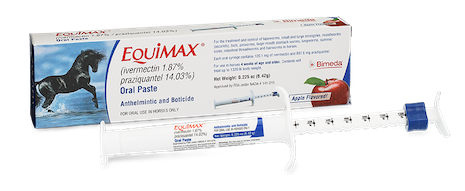
About Equimax
About EQUiMAX®
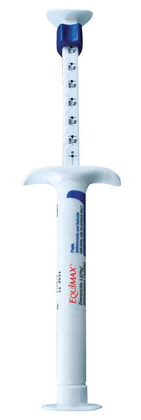
EQUiMAX® (ivermectin 1.87%/praziquantel 14.03%) is a broad spectrum parasite, bot and tapeworm control. It is safe to use in foals as early as 4 weeks of age, pregnant and lactating mares, and breeding stallions.
Indications
INDICATIONS
EQUiMAX® has broad-spectrum activity against a range of debilitating and performance-depriving parasites including:
- Tapeworms (Anoplocephala perfoliata)
- Large Strongyles (adults) (Strongylus vulgaris) also early forms in blood vessels), (S. edentatus) (also tissue stages), (S. equinus), (Triodontophorus spp.)
- Small Strongyles (adults, including those resistant to some benzimidazole class compounds) (Cyathostomum spp.), (Cylicocyclus spp.), (Cylicostephanus spp.), (Cylicodontophorus spp.)
- Small Strongyles (fourth–stage larvae)
- Pinworms (adults and fourth–stage larvae) (Oxyuris equi)
- Ascarids (adults and third– and fourth–stage larvae) (Parascaris equorum)
- Hairworms (adults) (Trichostrongylus axei)
- Large-mouth Stomach Worms (adults) (Habronema muscae)
- Bots (oral and gastric stages) (Gasterophilus spp.)
- Lungworms (adults and fourth–stage larvae) (Dictyocaulus arnfieldi)
- Intestinal Threadworms (adults) (Strongyloides westeri)
- Summer Sores caused by Habronema and Draschia spp. cutaneous third-stage larvae
- Dermatitis caused by Neck Threadworm microfilariae, Onchocerca sp.
Benefits
BENEFITS
- Apple flavored
- A smooth, quick-dissolving paste
- Broad spectrum activity against a wide range of parasites
- Proven safe in horses of all ages
- A single syringe contains enough active ingredients to treat horses weighing up to 1,320 lb
- Easy-to-use-and-handle syringe/applicator for more accurate dosing
Competitor Comparison
| EquiMax® | Other Available Brands | ||
|---|---|---|---|
| Active Ingredients | 1.87% ivermectin 14.03% praziquantel |
1.55% ivermectin 7.75% praziquantel |
2.0% moxidectin 12.5% praziquantel |
| Foals (minimum age) |
4 weeks | 2 months | 6 months |
| Breeding Stallions | Yes | No | No |
| Pregnant Mares | Yes | No | No |
| Lactating Mares | Yes | No | No |
| Syringe Dosage | Up to 1,320 lbs | Up to 1,250 lbs | Up to 1,500 lbs |
| Flavoring | Apple | No Flavor | No Flavor |
| EZE-Grip Syringe | Yes | No | No |
EZE-GRIP™ Syringe
Click the Spots!
Apple Flavoring
We all know that horses love apples!
We also know that receiving the full dose of any medicine is fundamental in getting the complete benefit of the treatment. It is particularly important for antiparasitic treatments, for which under-dosing may increase the chances of those parasites developing resistance to the active ingredient, rendering it less effective over time.
 Horses are much more likely to ingest the full dose of medicine if it’s flavored pleasantly. Apple flavored de-wormers (such as Equimax®) can increase the ease of administration and ensure the horse receives the proper, full dose. This means less spitting, less product waste, better efficacy. Not counting a more pleasant experience for both owner and horse.
Who knew something as simple as an apple flavor can go such a long way!
Available Pack Sizes
| Product Code | Unit Size | Case |
| 1EQU022 | 6.42g | 72 |
| 1EQU024 | 6.42g | 48 |
Documents To Download

For more information, please view the EQUIMAX® product information sheet.
Consult your Veterinarian to determine the best deworming program for your horse.
FOR ORAL USE IN HORSES 4 WEEKS OF AGE AND OLDER. EQUIMAX® (ivermectin/praziquantel) Paste should not be used in other animal species as severe adverse reactions, including fatalities in dogs, may result. Do not use in horses intended for human consumption. Swelling and itching reactions after treatment with ivermectin paste have occurred in horses carrying heavy infections of neck threadworm (Onchocerca sp. microfilariae), most likely due to microfilariae dying in large numbers. Not for use in humans. Ivermectin and ivermectin residues may adversely affect aquatic organisms, therefore dispose of product appropriately to avoid environmental contamination. Trademarks belong to their respective owners.